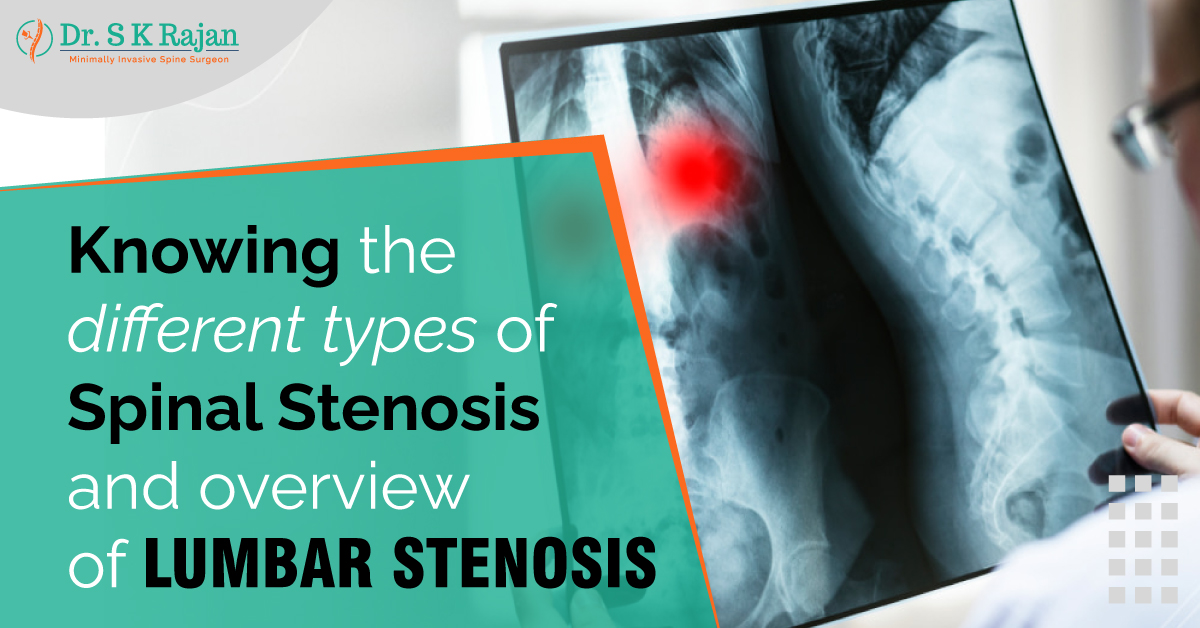
Spinal stenosis is any narrowing of the nerves that leave the spine, including the spinal cord and nerve roots. Several things, such as a herniated disc, an abnormal alignment, a bulging disc, scoliosis, or the growth of a bone spur, can cause the narrowing. The type of stenosis is based on where the stenosis is. There are three main types: lateral recess stenosis, central spinal stenosis, and foraminal stenosis. Each can happen in any part of the spine (i.e., cervical, thoracic, or lumbar).
Central stenosis is the narrowing of the space between the vertebrae in the middle of the spine, which is where the spinal cord goes. The spinal cord begins in the neck, moves down through the thoracic spine, and ends at the conus medullaris, about the second level down in the lumbar spine. Central canal stenosis is most common in the neck and lowers back because these spine parts move the most.
Foraminal stenosis is a narrowing of the foramen, the spaces between the vertebrae where nerves leave the spinal cord and go to other parts of the body. These give the legs, arms, and torso the ability to move and feel. Foraminal stenosis happens most often in the lumbar spine and cervical because these are the parts that move the most and the lumbar spine also supports the body's weight. This is the type of spinal stenosis that happens most often.
In lateral recess stenosis, the channels that nerves go through before they leave through the foramen get smaller. Before the nerve goes through the foramen, it starts to grow in the lateral recess right next to the spinal cord. This kind of stenosis affects the nerves that branch off the spinal cord and give the arms, legs, and torso their motor functions.
The lumbar spine is the part of the back between the ribs and the pelvis. It is made up of five vertebrae. Lumbar spinal stenosis is when the spinal canal gets smaller, putting pressure on the nerves from the lower back to the legs. It can happen to younger people because of how they develop, but it usually happens to people aged 60 and up because it is a degenerative disease.
Most of the time, the spinal canal gets smaller over many years or decades. Ageing makes the discs less spongy, which causes the discs to lose height and may cause the hardened disc to bulge into the spinal canal. There may also be bone spurs and thickening of the ligaments. All these can cause the central canal to narrow, which may or may not cause symptoms. The cause of the symptoms could be inflammation, pressure on the nerve(s), or both.
Some of these symptoms are pain, weakness, or numbness in the calves, legs, or buttocks; cramping in the calves when walking, which makes it hard to walk far without
stopping often; pain that spreads into one or both thighs and legs, which is similar to what people call "sciatica"; in rare cases, loss of motor function in the legs or bowel or bladder problems; pain that gets better when bending forward, sitting, or lying down.
Lumbar spinal stenosis can be caused by degenerative spondylolisthesis and degenerative scoliosis, a spine curvature. Osteoarthritis of the facet joints leads to degenerative spondylolisthesis when one vertebra slides over another. Most of the time, the L4 vertebra slides over the L5 vertebra. Most of the time, it is treated the same way as lumbar spinal stenosis: with conservative and surgical methods.
Most cases of degenerative scoliosis happen in the lower back, and people 65 and older are most likely to get it. Back pain from degenerative scoliosis usually starts slowly and gets worse when you do things. In this type of scoliosis, the spine usually only slightly curves. Surgery may be needed if nonsurgical treatments don't help ease the pain of the condition.
A neurosurgeon makes a diagnosis based on the patient's history, symptoms, physical exam, and test results.
The following imaging studies may be used: